
Human Eye Model — 5x Enlarged, 6-Part for Ophthalmology & Anatomy Teaching
Detailed institutional product information, specifications, use cases and quotation support from VistaStay Med.
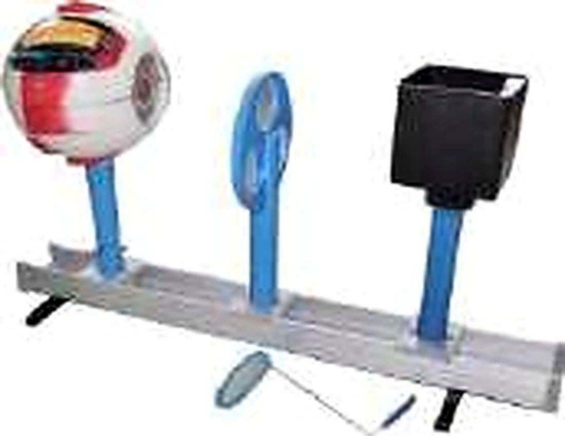
Human Eye Model — 5x Enlarged, 6-Part for Ophthalmology & Anatomy Teaching
5x enlarged 6-part human eye model showing retina, lens, cornea & optic nerve for medical colleges & nursing institutes. Detailed, durable. Pan-India delivery. Enquire now.
See every detail of the visual system. This 5x enlarged, 6-part eye model reveals the retina, lens, cornea, iris, ciliary body and optic nerve in extraordinary detail — making it perfect for ophthalmology teaching, anatomy practicals and patient education in medical colleges and eye hospitals.
Product Specifications
| Model Reference | REF-340 |
| Scale | 5x enlarged (approx. 15cm diameter) |
| Material | High-grade PVC |
| Parts | 6 parts: sclera, choroid, retina, lens, cornea, optic nerve |
| Structures Shown | Cornea, iris, pupil, lens, ciliary body, retina (with macula and fovea), optic disc, optic nerve, sclera, choroid, vitreous humour |
| Base | Plastic stand with bony orbit representation |
| Warranty | 1 year against manufacturing defects |
| Ideal For | MBBS anatomy, Ophthalmology, Nursing, Optometry |
Detailed Description
The VistaStay Med human eye model (REF-340) is a 5x enlarged, 6-part representation that brings ocular anatomy into stunning clarity. At approximately 15cm in diameter, every structure — from the transparent cornea to the optic nerve head — is visible and accessible for hands-on study.
The model disassembles into six components: the outer sclera (with removable cornea and iris), the choroid layer, the detailed retina (showing the macula, fovea and optic disc), the crystalline lens, the ciliary body with suspensory ligaments, and the optic nerve with cross-sectional detail. The bony orbit base provides anatomical context for the eye's position within the skull.
Students can trace the path of light from cornea → aqueous humour → pupil → lens → vitreous → retina → optic nerve, correlating structure with visual function. The enlarged scale makes this model equally valuable for ophthalmology residents reviewing surgical anatomy and for nursing students learning eye examination techniques.
Key Features
- ✅ 5x enlarged for extraordinary detail visibility
- ✅ 6-part disassembly: sclera, choroid, retina, lens, ciliary body, optic nerve
- ✅ Detailed retina with macula, fovea and optic disc
- ✅ Removable cornea and iris for anterior segment study
- ✅ Crystalline lens with realistic curvature
- ✅ Bony orbit base for anatomical context
- ✅ Hand-painted for realistic tissue differentiation
- ✅ Quality-checked and securely packaged for pan-India delivery
Use Cases
- 🏥 Medical college anatomy and ophthalmology practicals
- 🩺 Nursing institute eye examination training
- 👁️ Eye hospital patient education displays
- 🏫 Optometry college ocular anatomy teaching
- 📚 AYUSH college anatomy labs
Indicative Pricing
💰 Institutional Price Range: ₹3,200 – ₹5,000
(Varies by brand and quantity)